All year round, and particularly in summer, your skin is exposed to ultraviolet rays, which brings new brown patches, localised hyperpigmentation and new moles.
All year round, and particularly in summer, your skin is exposed to ultraviolet rays, which brings new brown patches, localised hyperpigmentation and new moles. These changes in the appearance of the skin should not be ignored, which is why an appointment with a dermatologist is highly advisable, especially if you have noticed new moles appearing.
Older moles you can examine yourself, looking at their shape, their size and their colour. The ABCDE rule is a great help in self-examination. Anyone can assess their own moles by following an algorithm that is extremely simple and at the same time effective, and that gives a warning to see a dermatologist promptly.
This algorithm, known in short as ABCDE, has been in use for a long time and is within anyone's reach where they have one or more naevi on the skin whose appearance needs watching over time.
If a mole shows one or more of the changes below, which may appear over varying periods of time, a specialist consultation at a dermatology clinic is called for:
• A – asymmetry, where one half of the naevus does not match the other half
• B – where the borders of the naevus are irregular, broken or indistinct
• C – where the mole examined shows different colours: different shades of brown, black or red areas, or white portions with no pigment
• D – where the diameter of the naevus is greater than 6 mm
• E – where part of the mole is elevated, raised above the surrounding skin.
If you notice major changes, or you have local symptoms (you can feel the mole on your skin), you should see a dermatologist, who will carry out a dermatoscopic examination of that lesion.
Dermatoscopy is a new, revolutionary method that helps in diagnosing, monitoring and distinguishing between pigmented lesions of the skin.
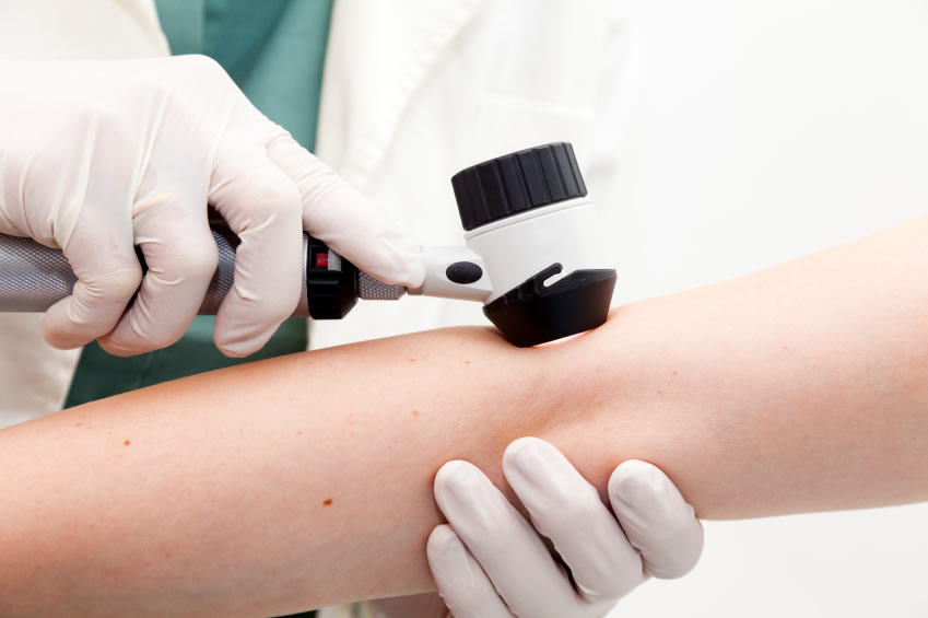
Dermatoscopy (also called dermoscopy or epiluminescence microscopy) is the examination of moles and other skin lesions using a special instrument called a dermatoscope. The dermatoscope, or dermoscope, is an instrument made up of an optical system resembling a lens, with a light source attached. The dermatologist places this instrument over the skin lesion to be examined, so that it can be analysed in detail. Digital dermoscopic photographs of the moles can also be captured, making up a dermoscopic map recorded on a CD. This map can be consulted periodically to analyse the changes that have occurred over time, after injury or after sun exposure, and is decisive in the decision to excise a lesion surgically.
Being able to examine the skin in detail helps guide the diagnosis of skin cancers, particularly the diagnosis of malignant melanoma and the distinction between malignant melanoma and ordinary moles. This makes it possible to detect malignant melanoma early, at a curable stage.
The method is non-invasive and relatively easy to carry out; it calls for no particular preparation on the patient's part and can bring additional peace of mind, especially for patients with many moles.
When is this method indicated?
People at higher risk of skin cancer should have a dermatoscopic examination once or twice a year. This group includes people:
• with fair skin, blond hair and blue eyes, and with numerous freckles or sun spots
• who burn easily in the sun
• who have numerous brown or black patches on the skin, of varying shape and size
• who have or have had relatives with skin cancer
• who have habitually spent time in the sun, have not used sunscreens, and have frequently been sunburnt.
A dermatoscopic check is also absolutely necessary for any skin lesion that:
• has appeared recently
• has grown rapidly
• has been accidentally broken, itches or bleeds
• has an irregular outline
• is uneven in colour, or has changed colour lately
• has changed appearance rapidly
This procedure is strongly indicated for people in the “at risk” group (people with a history of skin cancer, with numerous naevi, or with naevi that look dysplastic or unusual).
The most formidable enemy of people with moles on their skin is the sun. The harmful effects of solar radiation are due to ultraviolet A and B rays. UVA and UVB can set off damage at the cellular level, causing genetic changes in DNA that may lead to skin cancer.
These negative effects, in the short and the long term, can be avoided by using a product with high sun protection (SPF 50+) and an effective filtering system against UVA radiation.
UVB radiation is partly stopped by the epidermis. It stimulates tanning, but it is also mainly responsible for sunburn.
UVA radiation accounts for 95% of the UV radiation at the earth's surface. It is present even on cloudy days, from sunrise to sunset, and can pass through glass. UVA penetrates deeper into the skin, as far as the cells of the dermis. It accelerates the ageing of the skin and can cause photodermatoses (benign summer light eruption) and hyperpigmentation (the mask of pregnancy — melasma — and pigment spots).
Sunscreens are divided, according to the protection they provide (the value of the sun protection factor, SPF), into creams with very high, high, moderate and minimal protection.
Very high protection (SPF 50+) is needed for people with very fair skin, for skin that is hypersensitive to the sun, for those diagnosed with photodermatoses, and for exposure to strong sunlight (the seaside, high altitudes, the tropics).
High protection (SPF 40) is recommended for children, for fair skin prone to sunburn, and for periods of exposure to strong sun.
Moderate protection complements high protection, or suits darker skin. Minimal protection is indicated for skin that is already tanned, or for sunlight of ordinary intensity.