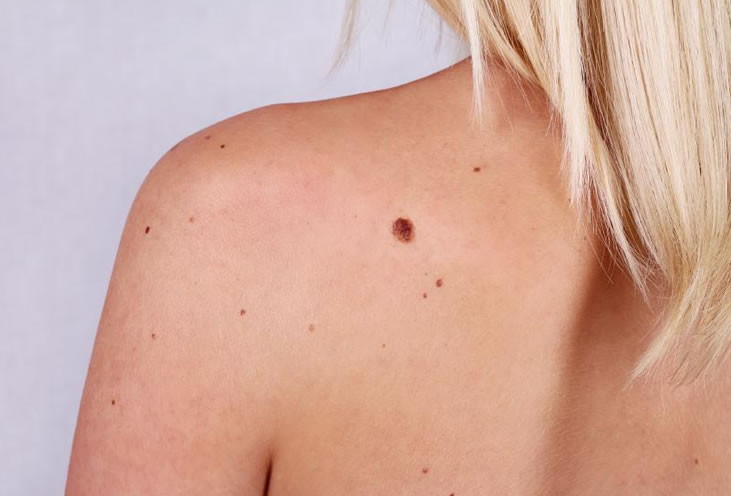
Moles are benign skin lesions arising from the proliferation of the cells that produce pigment. What they are, which kinds exist and how they are treated.
If you have several moles on your body and wonder whether you should have them checked by a dermatologist, or how to have them removed safely, you have come to the right place. We explain in turn what moles are, which kinds exist, when moles become dangerous and how to treat them properly.
What are moles (in scientific terms, naevi)?
A naevus (a mole) is a benign skin lesion arising from the proliferation of the cells that produce pigment (the melanocytes).
Naevi, or moles, may be present from birth (congenital naevi) or may appear later (acquired naevi). The risk of an ordinary mole turning into a melanoma is very small – about 1 in 3000 in men and 1 in 11,000 in women over a whole lifetime. Even so, regular monitoring remains essential.
Who can develop moles?
Most people have moles.
- 1% of the population is born with one or more congenital naevi
- people with fair skin tend to have more moles than people with dark skin
- the moles that appear in childhood (between the ages of 2 and 10) tend to be the most prominent and the most persistent over a lifetime
- the moles that appear later in life usually follow sun exposure
Why do moles appear?
Although the exact cause of the proliferation of the pigment-producing cells is not known, it is certain that the number of moles depends on genetic factors, on sun exposure and on immune status.
- People with many moles usually belong to families whose members have numerous moles, similar in appearance
- Somatic mutations in the RAS/RAF genes are associated with the appearance of congenital naevi
- New moles can appear after medicines that inhibit the BRAF genes (vemurafenib, dabrafenib)
- People chronically exposed to the sun (Australia, New Zealand) have more moles than their relatives living in less sunny places (northern Europe)
- Taking immunosuppressive medicines leads to an increase in the number of moles
What do moles look like clinically?
- They can appear anywhere on the body
- They may be flat or raised
- They can vary in colour from skin-coloured, pink, light or dark brown, to blue or black
- People with fair skin tend to have lighter moles, while those with dark skin have dark brown or black ones
- Although they are usually round or oval, they can also have unusual shapes
- They vary in size from a few millimetres to several centimetres across
Kinds of mole
By when they appear, moles fall into two broad categories: congenital (present at birth) and acquired (appearing over the course of life). Within each category there are several subtypes, differing in appearance and in risk.
Congenital moles – kinds and images
Congenital naevi appear at birth or in the first months of life and affect roughly 1 to 3% of newborns. They are classified by size:
- Small congenital naevus, under 1.5 cm across, on the skin of the arm
- Medium congenital naevus, between 1.5 and 19.9 cm, evenly pigmented
- Large congenital naevus, over 20 cm across
- Giant congenital naevus, over 40 cm, with an increased risk of melanoma
- Hairy congenital naevus, with thick hairs on its surface
- Café au lait macules – light brown patches on the skin
- Lentiginous naevus – a brown patch with darker dots
- Naevus of Ota – a brown-blue patch on the cheek and forehead
- Mongolian spot – a bluish patch over the lower back
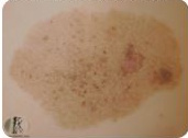
Lentiginous naevus
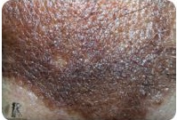
Giant congenital naevus
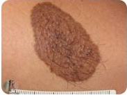
Medium congenital naevus
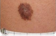
Small congenital naevus
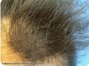
Hairy congenital naevus
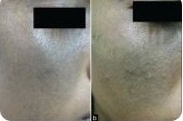
Naevus of Ota
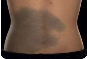
Mongolian spot
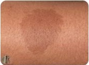
Café au lait macules
Acquired moles – kinds and images
Acquired naevi are the commonest and appear in childhood, in adolescence and in young adulthood. They are classified by the position of the melanocytes in the skin:
- Common naevus – a flat mole of a single colour, evenly distributed
- Common naevus on dark skin, dark brown or black
- Atypical naevus with irregular borders and uneven pigment, an irregular pattern with an increased risk of turning into melanoma
- Blue naevus – a dermal naevus with the pigment sitting deep in the skin
- Cellular naevus – an unpigmented, skin-coloured dermal naevus
- Miescher naevus – a soft, raised mole, common on the face
- Unna naevus – a papillomatous dermal naevus with an irregular surface
- Meyerson naevus – a mole surrounded by an eczematous halo
- Sutton naevus (halo naevus) – a mole with a white, depigmented halo around it
- Spitz naevus – a pink or brown mole in children and young people
- Reed naevus – a heavily pigmented, round mole, found on the limbs
- Recurrent naevus – pigment reappearing in the scar after a mole has been removed
- Agminated naevus – a cluster of small naevi or pigmented freckles
- Acral naevus – a mole on the palms or the soles
- Nail naevus – a mole appearing in the nail (a pigmented band)
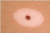
Sutton naevus (halo naevus)
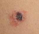
Meyerson naevus
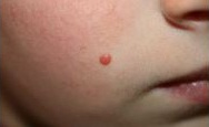
Spitz naevus
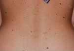
Common naevus on dark skin
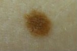
Common naevus
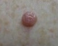
Cellular naevus
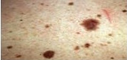
Atypical naevus with irregular borders
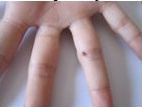
Acral naevus
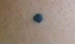
Blue naevus
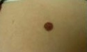
Unna naevus
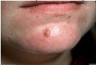
Miescher naevus
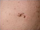
Agminated naevus
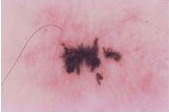
Recurrent naevus
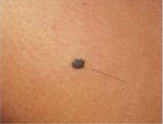
Reed naevus
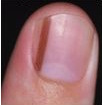
Nail naevus
Common moles versus atypical moles
| Common mole | Atypical mole | Possible melanoma |
| Round, symmetrical | Irregular | Asymmetrical |
| Clean, well defined | Faded, uneven | Ragged, indistinct |
| Uniform | Varied (2–3 shades) | Very varied (black, red) |
| Under 6 mm | 5–12 mm | Over 6 mm, growing (but can be smaller) |
| Stable | Slowly changing | Changing rapidly (weeks to months) |
| Very small | Increased (2 to 12 times) | Oncological risk |
What are the complications of moles?
People worry about moles because they have heard of melanoma, which is the malignant proliferation of melanocytes and the leading cause of death among skin cancers.
At the start, a melanoma can look like an ordinary mole, but over time its structure becomes disorganised and it can grow.
Moles can also change for other reasons unrelated to turning into melanoma, which nonetheless call for a dermatological consultation: sun exposure and pregnancy make them darker, and sometimes an eczematous halo may appear, reddish, dry and itchy (Meyerson naevus), or a white halo (Sutton naevus).
People with a large number of moles (over 100) are at greater risk of developing melanoma.
The ABCDE rule – how to recognise a suspicious mole
Use the ABCDE rule at home, once a month, to identify the suspicious moles that need a dermatological assessment:
- A – Asymmetry: one half of the mole does not look like the other half
- B – Border: the borders are irregular, ragged, indistinct or very abrupt
- C – Colour: the colour is not uniform and shows several shades (brown, black, red, blue, grey, white)
- D – Diameter: the mole is over 6 mm (the size of a pencil eraser)
- E – Evolution: the mole changes over time – it grows, changes shape or colour, or begins to itch, bleed or crust
If you notice one or more of the ABCDE signs, make an appointment with a dermatologist without delay.
A monthly self-examination guide – step by step
Set aside time once a month for a complete self-examination. Here is how to go about it properly:
- Stand in front of a large mirror in a well-lit room. Keep a small hand mirror nearby for the areas that are hard to see.
- Check the face, the neck, the ears and the scalp. On the scalp, use a comb to part the hair and see the skin.
- Examine the chest and the abdomen. Women should lift the breasts to see the skin beneath them.
- Check the upper arms, the underarms, the forearms, the palms, the backs of the hands and between the fingers.
- Carry on with the back of the body – use the hand mirror to see your back, your buttocks and the backs of your thighs.
- Move on to the legs – thighs, knees, calves, ankles, soles and between the toes. Do not forget the nails
- Check the intimate areas too, including the genital and perineal region.
When is a dermatoscopic consultation needed?
- When a mole changes size, shape, structure or colour
- When a new mole appears after the age of 40
- When it has the ABCDE features
- If it bleeds, crusts or becomes itchy
- If you have intense or prolonged sun exposure
- If you have a large number of moles (over 50)
- If you have a family history of melanoma or skin cancer
Most moles are benign on dermatoscopic assessment and are reassessed periodically where that is needed.
Moles that raise a suspicion of melanoma should be excised surgically and examined histopathologically.
How is a mole diagnosed?
Moles are generally diagnosed by clinical examination. If the moles are numerous or look unusual (atypical), the dermatologist will turn to dermatoscopic examination.
Digital dermatoscopic examination helps guide the diagnosis of skin cancers, particularly the diagnosis of malignant melanoma and the distinction between malignant melanoma and ordinary moles. This makes it possible to detect malignant melanoma and other skin cancers early, at a curable stage, and prevents unnecessary excisions.
The method is non-invasive and relatively easy to carry out, and calls for no particular preparation on the patient's part.
For those with a large number of moles, a digital dermatoscopic map is made, which involves recording and mapping every mole photographically, both macroscopically and dermoscopically, so that their evolution can be compared at later periodic examinations.
When, and for whom, is a dermatoscopic consultation needed?
- When a mole changes size, shape, structure or colour
- When a new mole appears (over 40)
- When it has the ABCD features (asymmetry, irregular borders, uneven colour, diameter over 6 mm)
- If it bleeds, crusts or becomes itchy
- If there has been intense or prolonged sun exposure
- If the moles are numerous
Most moles are benign on dermatoscopic assessment and are then reassessed periodically (where that is needed).
Moles that raise a suspicion of melanoma should be excised surgically and examined histopathologically.
What is the treatment for moles?
Most moles are harmless and can be left alone.
They may be removed in the following circumstances:
- To rule out skin cancer
- If they cause discomfort: irritated by clothing, a comb or a razor
- For cosmetic reasons: if they look unsightly
The surgical techniques include
- Surgical excision with histopathological examination of suspicious (atypical) naevi
- Destruction by radio- or electrosurgery (only in certain cases, with histopathological examination, on the doctor's instructions)
- CO2 laser vaporisation (only in certain cases, on the doctor's instructions, with histopathological examination)
Can moles be prevented from appearing?
The number of naevi can be kept down by strict sun protection, from birth onwards.
Sun protection through creams alone is not enough to prevent new naevi from appearing; wearing a hat and long-sleeved tops in sunny places is also recommended.
At any age, sun protection is needed to reduce the premature ageing of the skin and to lower the risk of skin cancer.
How to protect your skin properly
- Apply an SPF 50+ cream daily to every exposed area, even on cloudy days
- Reapply the cream every 2 hours when you are at the beach or playing sport outdoors
- Avoid the sun between 11:00 and 16:00, when UV radiation is at its strongest
- Wear a wide-brimmed hat, sunglasses and long sleeves in strong sun
- Do not use sunbeds – they significantly increase the risk of melanoma
- Protect children particularly: sunburn in childhood increases the risk of melanoma in adult life
Moles and pregnancy – what you should know
During pregnancy, hormonal changes can make existing moles darker or make them grow slightly. In most cases this is normal. If you notice rapid or asymmetrical changes, though, or moles developing the ABCDE features, make an appointment with a dermatologist. Dermatoscopic examination is safe in pregnancy.
Moles in children
Children develop moles mainly between the ages of 2 and 10, and these tend to be the most stable and persistent over a lifetime. It is normal for new moles to appear in children, but rapid changes should be assessed by a paediatric dermatologist.
Spitz naevi are common in children and can look alarming (pink-red, growing quickly), but they are benign. The correct diagnosis is made only by dermatoscopy and, where necessary, biopsy.
Particular attention is due to giant congenital naevi – these need monitoring by a multidisciplinary team (dermatologist, paediatric surgeon).
What is the outlook with naevi?
Most naevi appearing in childhood remain indefinitely. Adolescents and young adults tend to have the greatest number of naevi. As we grow older the moles become fewer, since they tend to disappear by themselves.
To improve the chance of detecting a melanoma at an early stage, the following are recommended:
- Self-examination of the skin every month
- Seeing a dermatologist where there are significant changes in the naevi or where new lesions appear,
- Periodic dermatoscopic examination where there are large numbers of moles, atypical naevi, or a personal or family history of skin cancer
- A digital dermatoscopic map, for patients at increased risk of melanoma, particularly those with many naevi
What counts as an increased risk of melanoma, or when moles become dangerous?
- A history of dysplastic naevi and melanoma
- A newly appeared naevus, or one that has changed shape, size or appearance
- Having more than 50 naevi, 20 of them over 2 mm across
- Melanoma in the family history
- Skin showing signs of sun damage (patches, freckles)
- Fair skin or eyes
- Intense and prolonged sun exposure
- Immunosuppression
Telling moles apart from other similar lesions
Many people confuse moles with other skin lesions that are not naevi. Here are the commonest confusions:
- Freckles (ephelides) – small, flat, light brown patches that come and go with sun exposure. They are not moles and carry no cancer risk.
- Solar lentigines – flat brown patches appearing after prolonged sun exposure, often on the hands, the face and the décolletage in people over 40. Benign, but a sign of “sun ageing”.
- Seborrhoeic warts (seborrhoeic keratoses) – raised brown or black lesions with a “stuck on” appearance. Benign, common after the age of 50.
- Viral papillomas – small, soft growths in areas of friction (neck, underarms, eyelids). Benign.
- Haemangiomas – red or purple raised lesions made up of blood vessels. Benign.
Only the dermatologist, through clinical and dermatoscopic examination, can tell them apart correctly.
Frequently asked questions about moles
Is it dangerous to have a mole removed?
It is not dangerous if the procedure is carried out by a dermatologist after a prior dermatoscopic assessment. Removing a mole does not cause melanoma, as some people believe. The real risk is removing an atypical mole without a biopsy and so delaying the diagnosis of a melanoma.
Can I remove moles at home with natural methods?
No. Never try to remove moles with over-the-counter solutions, plants, vinegar or other “natural” methods. You risk infections, ugly scars and, more seriously, losing the chance of a correct diagnosis if the mole was an early melanoma.
How often should I see a dermatologist for a check?
If you have fewer than 20 moles and no changes, once a year is enough. If you have more than 50 moles, atypical naevi or a family history of melanoma, have checks every 6 months and a digital dermatoscopic map every year.
Can moles come back after I have them removed?
If the mole was excised correctly and completely, it does not come back. With laser or radiofrequency removal there is a small risk of recurrence if residual cells remain. This is why careful assessment before the procedure is essential.
How long does recovery take after a mole is removed?
Laser or radiofrequency: 7 to 14 days for the crust to heal, and the skin regains its normal colour in 1 to 3 months.
Surgical excision: the stitches come out at 7 to 14 days (depending on the area), and the scar matures over 6 to 12 months.
My mole is growing. Does that mean it is cancer?
Not necessarily. Moles grow and change naturally in childhood, in adolescence and in pregnancy. What should alarm you is rapid growth (over weeks or a few months), asymmetry, irregular borders or varied colour – the ABCDE signs. If any changes appear, make an appointment with a dermatologist.
How much does it cost to have a mole removed?
The cost depends on the number and kind of moles, the method used, and whether histopathological examination is needed. For the current prices at Dermestetica, see the prices page or call 0733 297 207.
Bibliography
American Academy of Dermatology – Moles: Overview: aad.org
DermNet NZ – Melanocytic Naevus: dermnetnz.org/topics/melanocytic-naevus
National Cancer Institute – Common Moles, Dysplastic Nevi, and Risk of Melanoma: cancer.gov
Skin Cancer Foundation – The ABCDEs of Melanoma: skincancer.org
Mayo Clinic – Moles: Symptoms and Causes: mayoclinic.org